Citation
A.R. Harris, L. Peter, J. Bellis, B. Baum, A.J. Kabla, and G.T. Charras
PNAS 109:16449-16454 (2012)
Abstract
Abstract
One-cell-thick monolayers are the simplest tissues in multicellular organisms, yet they fulfill critical roles in development and normal physiology. In early development, embryonic morphogenesis results largely from monolayer rearrangement and deformation due to internally generated forces. Later, monolayers act as physical barriers separating the internal environment from the exterior and must withstand externally applied forces. Though resisting and generating mechanical forces is an essential part of monolayer function, simple experimental methods to characterize monolayer mechanical properties are lacking. Here, we describe a system for tensile testing of freely suspended cultured monolayers that enables the examination of their mechanical behavior at multi-, uni-, and subcellular scales. Using this system, we provide measurements of monolayer elasticity and show that this is two orders of magnitude larger than the elasticity of their isolated cellular components. Monolayers could withstand more than a doubling in length before failing through rupture of intercellular junctions. Measurement of stress at fracture enabled a first estimation of the average force needed to separate cells within truly mature monolayers, approximately ninefold larger than measured in pairs of isolated cells. As in single cells, monolayer mechanical properties were strongly dependent on the integrity of the actin cytoskeleton, myosin, and intercellular adhesions interfacing adjacent cells. High magnification imaging revealed that keratin filaments became progressively stretched during extension, suggesting they participate in monolayer mechanics. This multiscale study of monolayer response to deformation enabled by our device provides the first quantitative investigation of the link between monolayer biology and mechanics.
Figure sample
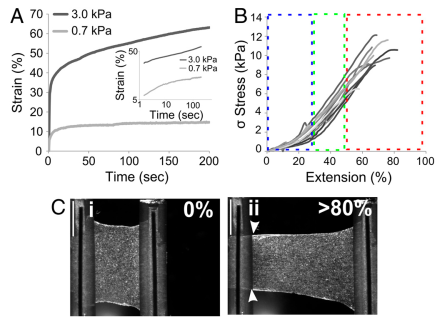
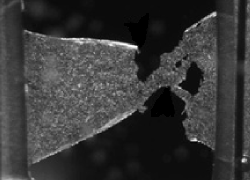
Mechanical properties of cell monolayers. (A) Creep response follow- ing step application of low (0.7 kPa, grey) and high (3 kPa, black) stress. The plotted responses are averaged over at least six experiments each. At low stress, following a rapid increase in strain, monolayers reached a plateau that lasted for the remainder of the experiment. At high stress, no plateau was reached and strain increased continually with time. (Inset) Creep response curves plotted in log–log scales. The creep response at high stress (black) was well fitted by a linear function with slope β = 0.15 +/- 0.03; whereas at low stress the creep response was not linear. (B) Stress-extension curves shown for 12 different monolayers. All curves displayed three distinct regimes of loading: (i) an initial toe region (blue box) as the monolayer becomes loaded under tension, (ii) a linear extension regime (green box) from which an elastic modulus can be calculated, and (iii) a plateau (red box) which cor- responds to plastic deformation and eventual failure. (C) Deformation of a monolayer under stretch. Images acquired by bright-field microscopy for a monolayer at 0 and >80% extension. At >80% extension, the monolayer delaminated from the test rods (arrows) suggesting that cell–cell adhesion is stronger than cell–substrate adhesion for this geometry. (sb = 1 mm)