Citation
J. Fouchard, T.P.J. Wyatt, A. Proag, A. Lisica, N. Khalilgharibi, P. Recho, M. Suzanne, A. Kabla, G. Charras
PNAS 117(17):9377-9383 (2020)
Abstract
Significance
Abstract
Significance
Epithelial monolayers are sheet-like tissues composed of cells connected to one another that line the surface of all organs. These tissues change shape and bend during developmental morphogenesis or early in tumor formation. Yet, the amplitude of bending forces and how they integrate with tensile and compressive forces within the plane of the tissue remain largely unknown. By revealing the ability of epithelial monolayers to curl, we demonstrate that the polarization of contractile molecular motors within the tissue thickness generates high spontaneous curvature of the sheet. We quantify the corresponding torques and show that stretch and compression within the tissue plane can substantially impact curling. Such mechanical coupling could be at play during epithelial morphogenesis.
Summary
Epithelial monolayers are two-dimensional cell sheets which compartmentalize the body and organs of multicellular organisms. Their morphogenesis during development or pathology results from patterned endogenous and exogenous forces and their interplay with tissue mechanical properties. In particular, bending of epithelia is thought to result from active torques generated by the polarization of myosin motors along their apicobasal axis. However, the contribution of these out-of-plane forces to morphogenesis remains challenging to evaluate because of the lack of direct mechanical measurement. Here we use epithelial curling to characterize the out-of-plane mechanics of epithelial monolayers. We find that curls of high curvature form spontaneously at the free edge of epithelial monolayers devoid of substrate in vivo and in vitro. Curling originates from an enrichment of myosin in the basal domain that generates an active spontaneous curvature. By measuring the force necessary to flatten curls, we can then estimate the active torques and the bending modulus of the tissue. Finally, we show that the extent of curling is controlled by the interplay between in-plane and out-of-plane stresses in the monolayer. Such mechanical coupling emphasizes a possible role for in-plane stresses in shaping epithelia during morphogenesis.
Figure sample
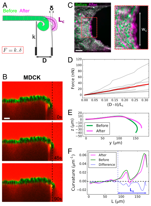
Characterization of active torques and bending modulus of epithelial monolayers. (A) Diagram of the setup to measure the bending modulus of an epithelial monolayer. A flexible glass capillary of stiffness k, serving as a force cantilever, is approached close to a tissue edge (green) of curled length L c . The displacement D imposed at the base of the cantilever generates a deflection δ of the cantilever, due to the restoring force of the flattened tissue (pink). (B) Time series of an MDCK monolayer imaged in profile during a ramp of displacement of the force cantilever. Dextran- Alexa647 was added to the medium (red). Note that this dye also stains the tip of the cantilever underneath the monolayer, which facilitates the measurement of its displacement. Cell membranes are marked with Cell- Mask (green). (Scale bar: 30 μm.) (C) Overlay of projected confocal stacks showing the free edge of an MDCK suspended monolayer before (green) and after (magenta) unfurling of the monolayer by the force cantilever. wc denotes the width of unfurled tissue. (Scale bar: 50 μm.) Inset shows a zoom on the deformed region of width wc. (Scale bar: 20 μm.) (D) Force variation along a ramp of displacement imposed at the cantilever base. The displace- ment of the cantilever tip is quantified by the ratio (D−δ)/ Lc , which is equal to 1 when the tissue is fully unfurled. The gray lines represent n = 9 separate experiments; the red line represents the average. (E) Typical profile of monolayer before (green) and after (pink) unfurling of the tissue by the cantilever. (F) Typical variation of curvature of the monolayer along its contour length L before (green) and after (pink) unfolding by the cantilever. The difference between the two curves is plotted in blue. The dashed blue line indicates the transition between curled and non-curled regions of the tissue corresponding to the position where the blue curve departs from the x axis.