
Citation
S. Jain, V.M.L. Cachoux, G.H.N.S. Narayana, S. de Beco, J. D’Alessandro, V. Cellerin, T. Chen, M.L. Heuzé, P. Marcq, R.-M. Mège, A.J. Kabla, C.T. Lim and B. Ladoux
Nature Physics (2020)
Abstract
Abstract
The directed migration of cell collectives is essential in various physiological processes, such as epiboly, intestinal epithelial turnover, and convergent extension during morphogenesis, as well as during pathological events such as wound healing and cancer metastasis. Collective cell migration leads to the emergence of coordinated movements over multiple cells. Our current understanding emphasizes that these movements are mainly driven by large-scale transmission of signals through adherens junctions. In this study, we show that collective movements of epithelial cells can be triggered by polarity signals at the single-cell level through the establishment of coordinated lamellipodial protrusions. We designed a minimalistic model system to generate one-dimensional epithelial trains confined in ring-shaped patterns that recapitulate rotational movements observed in vitro in cellular monolayers and in vivo in genital or follicular cell rotation. Using our system, we demonstrated that cells follow coordinated rotational movements after establishing directed Rac1-dependent polarity over the entire monolayer. Our experimental and numerical approaches show that the maintenance of coordinated migration requires the acquisition of a front–rear polarity within each single cell but does not require the maintenance of cell–cell junctions. Taken together, these unexpected findings demonstrate that collective cell dynamics in closed environments as observed in multiple in vitro and in vivo situations can arise from single-cell behaviour through a sustained memory of cell polarity.
Figure sample
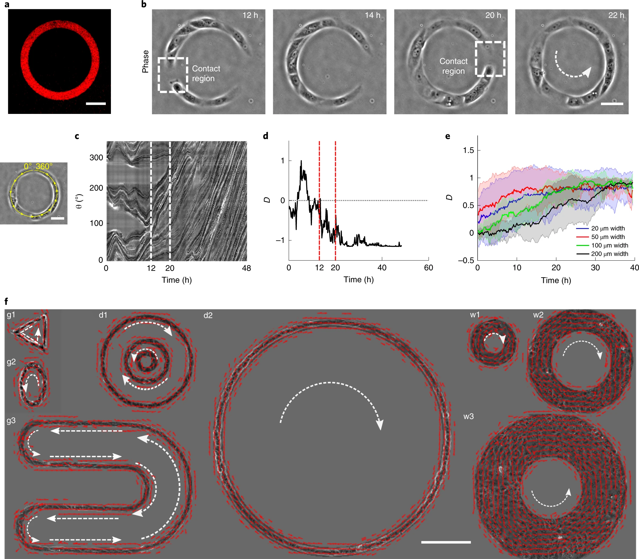
a, Microcontact-printed pattern of Cy3-conjugated fibronectin in the shape of an annular ring (ring outer diameter 200 µm, track width 20 µm). b, Phase contrast images of a train of MDCK cells after initial seeding and spreading. The dashed white box indicates the contact regions where small cell trains merged into larger trains at 12 h and 20 h. c, Spatio-temporal displacement of cells traced along the circumferential mid-line of the ring track. A small train-merging event is indicated by the white dashed lines. d, Evolution of coordination parameter, D, of cell trains in the ring with time (at t = 20 h the cell train in the ring begins to rotate in an anticlockwise direction). Time points with a small train-merging event are indicated by red dashed lines. e, D of cells in rings with different widths, 20 µm (total number of data points, n = 62; number of experiments, m = 3), 50 µm (n = 27, m = 2), 100 µm (n = 8) and 200 µm (n = 8), shows a collective migration of cell trains independent of track width. f, Velocity vectors representing directed collective cell migrations for a variety of confined geometries (g1—triangle, g2—ellipse, g3—U shape), diameters (d1, outer—400 µm, middle—200 µm, inner—100 µm; d2, 1 mm) and widths (w1—50 µm, w2–100 µm, w3—200 µm). All error bars indicated are s.d. Scale bars in a,b,c, 50 µm, and f, 200 µm.