
Citation
M. G. Linguraru, A. Kabla, N. V. Vasilyev, P. J. del Nido, R. D. Howe
Academic Radiology 14:1298 (2007)
Abstract
Rationale and Objectives
Abstract
Rationale and Objectives
Real-time cardiac ultrasound (US) allows monitoring the heart motion during intracardiac beating heart procedures. Our application assists pediatric atrial septal defect (ASD) closure techniques using real-time 3D US guidance and rigid instruments. ASD tracking is also an important tool for facilitating systematic clinical studies of the dynamic behavior of the intra-atrial communication. One major image processing challenge is associated with the required processing of information at high frame rate, especially given the low image quality.
Materials and Methods
We present an optimization scheme for a block flow technique, which combines the probability-based velocity computation for an entire block (a 3D volume centered on the ASD) with cyclic template matching. The adapted similarity imposes constraints both locally (from frame to frame) to conserve energy, and globally (from a reference template) to minimize cumulative errors. The algorithm is optimized for fast and reliable results. For tests, we use three intra-operational 4D ultrasound sequences of clinical infant beating hearts with ASD.
Results
Computing velocity at the block level with an optimized scheme, our technique tracks ASD motion at a frequency of 60 frames/s on clinical 4D datasets. Results are stable and accurate for changes in resolution and block size. In particular, we show robust real-time tracking and preliminary segmentation results of the ASD shape, size and orientation as a function of time.
Conclusions
We present an optimized block flow technique for real-time tracking of ASD to assist in minimally invasive beating heart surgery. Our method proposes the standard use of references for processing repetitive data. This paper represents, to our knowledge, the first study on the dynamic morphology of ASD that takes into account the angular effect introduced by the slanted position of the intra-atrial communication with respect to the US probe.
Figure sample
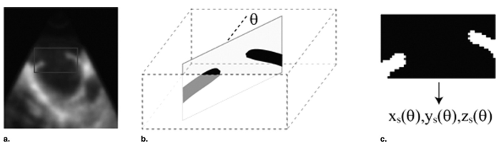
Atrial septal defect (ASD) segmentation: (a) a two-dimensional slice of the three-dimensional image in (x,z) coordinates, nor- mal to the ASD; the frame represents the typical block size and location used for the segmentation; (b) the neighborhood of ASD is ex- tracted using the tracking information; a family of vertical slices defined by the angle θ between the normal to the plane and the x axis are used to detect the boundary of the hole; (c) a typical vertical section after thresholding; the computed location of the hole boundary allows the reconstruction of the ASD shape parameterized by the angle θ.