
Citation
S. McDermott, J. Kim, A.A. Leledaki, D. Parry, L. Lee, A. Kabla, C. Mkindi, R. Bowman and P. Cicuta
Rev Sci Instrum 93:014104 (2022)
Abstract
Abstract
The process of making blood smears is common in both research and clinical settings for investigating the health of blood cells and the presence of blood-borne parasites. It is very often carried out manually. We focus here on smears for malaria diagnosis and research, which are frequently analyzed by optical microscopy and require a high quality. Automating the smear preparation promises to increase throughput and to improve the quality and consistency of the smears. We present here two devices (manual and motorized) designed to aid in the making of blood smears. These are fully documented, open-source hardware, and an important principle was to make them easily fabricated locally anywhere. Designs and assembly instructions are freely available under an open license. We also describe an image analysis pipeline for characterizing the quality of smears and use it to optimize the settings and tunable parameters in the two devices. The devices perform as well as expert human operators while not requiring a trained operator and offering potential advantages in reproducibility and standardization across facilities.
Figure sample
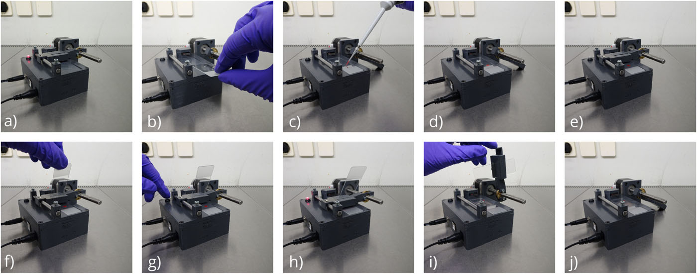
Operation of autohaem smear+ only requires the push of a button once the slides and the blood drop have been loaded. The device performs very reproducibly and autonomously, so higher overall throughput can be achieved by preparing the blood while the device operates or by having multiple devices running simultaneously. The sequence of images shows the key steps in the operation of the electro-mechanical device. (a) Plugging in the device, in which the device performs calibration. (b) Rotating the slider and placing the microscope slide in the slot. (c) and (d) Placing a drop of blood on the microscope slide. (e) Rotating the slider into position. (f) Inserting the spreader microscope slide. (g) Pressing the button to start the smear. (h) Device is producing the smear. (i) Removing the spreader slide.