
Citation
Y. Wang, K. Soga, J.T. Dejong and A.J. Kabla
Journal of Geotechnique and Geoenvironmental Engineering 145(9):04019045 (2019)
Abstract
Abstract
Microbial-induced calcium carbonate (CaCO₃) precipitation (MICP) has been explored for its potential engineering applications such as soil stabilization, but current understanding of the fundamental MICP processes at the microscale is limited. In this study, real-time in situ microscale experiments were conducted using glass slides and microfluidic chips (synthetic porous media that simulate soil matrices to model the conditions similar to actual MICP treatments) to visualize the CaCO₃ precipitation process. The results of this study show that irregularly shaped CaCO₃ precipitates initially emerged on bacterial aggregates and subsequently dissolved with time as regularly shaped CaCO₃ crystals started growing; less stable and smaller CaCO3 crystals may dissolve at the expense of growth of more stable and larger CaCO₃ crystals. The time-dependent phase transformation of CaCO₃ precipitates makes the size of the crystals formed during MICP highly dependent on the time interval between cementation solution injections during a staged-injection procedure. When the injection interval was 3–5 h, a larger number of crystals (200–1,000 per 10 6 μm 3 ) with smaller sizes (5–10 μm) was produced. When the injection interval was longer (23–25 h), the crystals were larger (10–80 μm) and fewer in number (5–20 per 10 6 μm³). The direct observation of MICP processes in this study improves the understanding of MICP fundamentals and the effect of MICP processes on the properties of CaCO₃ crystals formed after MICP treatment. These observations will therefore be useful for designing future MICP treatment protocols that improve the properties and sustainability of MICP-treated samples.
Figure sample
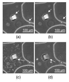
Microscope images of a pore in the microfluidic chip after the second injection of cementation solution: (a) 0 h; (b) 1 h; (c) 3 h; and (d) 24 h.