
Citation
J.P. Sharkey, D.C.W. Foo, A. Kabla, J.J. Baumberg and R.W. Bowman
Review of Scientific Instruments 87:025104 (2016)
Abstract
Abstract
Open source hardware has the potential to revolutionise the way we build scientific instruments; with the advent of readily available 3D printers, mechanical designs can now be shared, improved, and replicated faster and more easily than ever before. However, printed parts are typically plastic and often perform poorly compared to traditionally machined mechanisms. We have overcome many of the limitations of 3D printed mechanisms by exploiting the compliance of the plastic to produce a monolithic 3D printed flexure translation stage, capable of sub-micron-scale motion over a range of 8 × 8 × 4 mm. This requires minimal post-print clean-up and can be automated with readily available stepper motors. The resulting plastic composite structure is very stiff and exhibits remarkably low drift, moving less than 20 μm over the course of a week, without temperature stabilisation. This enables us to construct a miniature microscope with excellent mechanical stability, perfect for time-lapse measurements in situ in an incubator or fume hood. The ease of manufacture lends itself to use in containment facilities where disposability is advantageous and to experiments requiring many microscopes in parallel. High performance mechanisms based on printed flexures need not be limited to microscopy, and we anticipate their use in other devices both within the laboratory and beyond.
Figure sample
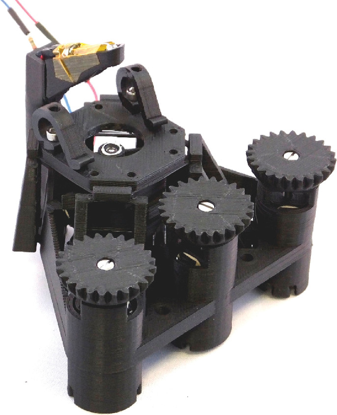
Photograph of the microscope. The three gears at the front control lateral motion (outer gears) and focus (centre), while the sample is held on the translating stage by two printed clips. A white LED is mounted on a printed arm at the top, and the lens (from the Raspberry Pi camera module) is visible through the hole in the sample stage. The camera sensor mounts underneath the microscope.